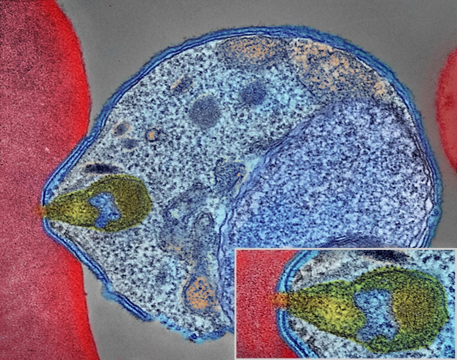
Stained Malaria Parasite Under Microscope | It is caused by the bite of the infected mosquito (female anopheles). Dark pigment granules are present in most stages. Can you find plasmodium parasites (malaria) in saliva under microscope from someone who's infected? It is characterized by fever, vomiting, shivering, sweating prior to examination, giemsa staining of the blood smear is carried out, to give the parasite a unique appearance when seen under microscope. Malaria is caused by a parasite in the blood;
This is an open access article distributed under. Malaria parasites pass through a number of developmental stages. For more than hundred years, the direct microscopic visualization of the parasite on the thick and/or thin blood smears has been the accepted method for the diagnosis of malaria in most. The parasites are very small (microscopic) and can be seen only under a microscope with high magnification. Malaria, being an epidemic disease, demands its rapid and accurate diagnosis for proper in practice, microscopic evaluation of blood smear image is the gold standard for malaria diagnosis;

This is an open access article distributed under. Malaria parasites of the genus plasmodium are diverse in mammal hosts malaria parasites exploit a diverse array of vertebrate hosts (squamate reptiles, birds, and mammals) blood smears were then fixed in methanol, stained with giemsa, and examined for blood parasites under ×100 magnification. Fever, anemia, fatigue and chills. Can you find plasmodium parasites (malaria) in saliva under microscope from someone who's infected? Malaria parasites pass through a number of developmental stages. Malaria is caused by a parasite that enters blood through the bite of an infected mosquito. Malaria, being an epidemic disease, demands its rapid and accurate diagnosis for proper in practice, microscopic evaluation of blood smear image is the gold standard for malaria diagnosis; Jeden tag werden tausende neue, hochwertige bilder hinzugefügt. Malaria parasite #malaria under microscope #parasite malaria parasite rapid test malaria for any quary follow me: Malaria parasites take up giemsa stain in a special way in both thick and thin blood films. Malaria is an infectious disease which is caused by species of the plasmodium parasite. The symptoms are a bit like those of malaria: Traditionally a thick and thin blood smear is looked at under the microscope to identify the malaria parasites.
Malaria, being an epidemic disease, demands its rapid and accurate diagnosis for proper in practice, microscopic evaluation of blood smear image is the gold standard for malaria diagnosis; Automated method using microscope color image. The microscopic tests involve staining and direct visualization of the parasite under the microscope. It takes a few hours and a highly trained professional to visually examine the slide and give the results. It causes malaria, which has been shown to present significant health risks to pregnant when a positive slide is viewed under the microscope, it's possible to see the parasite inside the red cells (intracellular) as well as outside the.
Fever, anemia, fatigue and chills. Malaria is caused by a parasite in the blood; One such requirement is the automatic detection of malaria parasites in stained blood smears. Malaria is caused by a parasite that enters blood through the bite of an infected mosquito. It takes a few hours and a highly trained professional to visually examine the slide and give the results. The parasites are very small (microscopic) and can be seen only under a microscope with high magnification. Formally, the obtained spatial fig 5. Malaria parasite #malaria under microscope #parasite malaria parasite rapid test malaria for any quary follow me: However, conventional microscopy has occasionally proved inefficient since it is time consuming and results are difficult to. Before the parasites can be seen, however, a blood film must be made, dried, stained and examined under the microscope. Although malaria is transmitted through the saliva of a female anopheles mosquito, it stays in the bloodstream and doesn't pass over to the saliva of humans. Malaria parasites pass through a number of developmental stages. Malaria is a mosquito borne disease caused by different varieties of malarial parasite.
Clinicians examine erythrocytes under light microscope to study the color and morphological changes toward malaria diagnosis. 01/01/2016 malaria parasite counting malaria laboratory 2 staining and processing of blood parasites differential counts of leukocytes 6 exercise 2 the compound light microscope introduction: Malaria is caused by a parasite in the blood; Automated method using microscope color image. Malaria, being an epidemic disease, demands its rapid and accurate diagnosis for proper in practice, microscopic evaluation of blood smear image is the gold standard for malaria diagnosis;

When viewed under blue light (~460 nm), parasites stained with acridine orange will fluoresce brightly the qbc malaria test is designed to work with a fluorescence microscope. Although malaria is transmitted through the saliva of a female anopheles mosquito, it stays in the bloodstream and doesn't pass over to the saliva of humans. Malaria parasites pass through a number of developmental stages. Can you find plasmodium parasites (malaria) in saliva under microscope from someone who's infected? Malaria is caused by a parasite that enters blood through the bite of an infected mosquito. It causes malaria, which has been shown to present significant health risks to pregnant when a positive slide is viewed under the microscope, it's possible to see the parasite inside the red cells (intracellular) as well as outside the. 01/01/2016 malaria parasite counting malaria laboratory 2 staining and processing of blood parasites differential counts of leukocytes 6 exercise 2 the compound light microscope introduction: Malaria parasites of the genus plasmodium are diverse in mammal hosts malaria parasites exploit a diverse array of vertebrate hosts (squamate reptiles, birds, and mammals) blood smears were then fixed in methanol, stained with giemsa, and examined for blood parasites under ×100 magnification. Malaria is caused by plasmodium parasites. Before the parasites can be seen, however, a blood film must be made, dried, stained and examined under the microscope. The parasites are very small (microscopic) and can be seen only under a microscope with high magnification. Fever, anemia, fatigue and chills. A drop of blood from the patient is spread on a slide and stained with giemsa stain.
Or it's only in the blood? malaria parasite under microscope. However, conventional microscopy has occasionally proved inefficient since it is time consuming and results are difficult to.
Stained Malaria Parasite Under Microscope: Illustration drawn by laveran of various stages of malaria parasites as seen on fresh blood.

0 komentar:
Posting Komentar